
SNB-19 (U-251MG derivative) xenograft model
NOTE: According to ATCC the analysis of short tandem repeats (STRs) indicated that the SNB-19 human glioblastoma cell line exhibits an STR pattern that is indistinguishable from that of the U-373 MG cell line (shown to be a U251MG derivative). Furthermore, SNB-19 and U-373 MG also demonstrate a commonality in their derivative chromosomes. These findings were validated using the original cell stocks held by the American Type Culture Collection (ATCC). According to Cellosaurus SNB-19 cell line has been shown to be a U-251MG derivative.
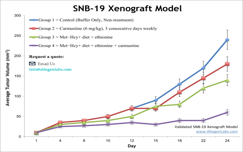
Prior to recent characterization of SNB-19 cell line (see NOTE above), this cell line was reported as derived from a glioblastoma multiforme, a highly malignant brain tumor. The American Brain Tumor Association has reported the complete lack of response for select glioblastoma types to treatment. The SNB-19 cell line was utilized in biological research related to human glioblastoma. In a 1988 study, published in Cancer Research Journal, the SNB-19 cell line was the most clonogenic in soft agar and showed the most tumorigenicity in nude mice. Bichat et al. (2000) reported in Clinical Cancer Research the enhanced efficacy of cytotoxic agents with simultaneous methionine depletion especially for treatment of drug-resistant tumors. In this study SNB-19 cells were used as a glioma xenograft model where survival duration was successfully extended with this regimen. A 2015 study (Latocha et al.) used in vitro SNB-19 cells to study the mechanism of action of phenothiazine amine derivatives; they looked at cytotoxic activity, apoptosis induction, effect on proliferation, cell cycle gene expression and total cell oxidative status. SNB-19 cells were used to create the SNB19 (U-251MG derivative) xenograft mouse model.
SNB19 Xenograft Model: Download ![]()
Download Altogen Labs SNB19 Xenograft Model PowerPoint Presentation: ![]()
SNB-19 (U-251MG derivative) Subcutaneous Xenografts in Preclinical Glioblastoma Research
Subcutaneous xenograft transplantation using SNB-19 (U-251MG derivative) cells serves as a foundational model in preclinical oncology, enabling reproducible and accessible evaluation of tumor growth and therapeutic response. The SNB-19 cell line, derived from human glioblastoma multiforme, exhibits key molecular alterations including PTEN loss, p53 dysfunction, and moderate EGFR expression, making it a clinically relevant tool for glioma research. Although lacking the native brain microenvironment, subcutaneous implantation allows for precise tumor measurement, efficient monitoring, and high-throughput compound screening. Studies utilizing SNB-19 xenografts have demonstrated the efficacy of DNA-damaging agents, histone deacetylase inhibitors, and nanoparticle-based therapeutics, revealing alterations in apoptotic and epigenetic pathways. Our research has optimized this model to ensure consistent tumor engraftment and growth kinetics, supporting its use for early-phase drug evaluation, biomarker discovery, and downstream molecular analyses. While orthotopic models offer greater physiological relevance, the SNB-19 subcutaneous xenograft remains an indispensable system for translational glioblastoma studies.
Modeling Intracranial Glioma Progression
Orthotopic xenograft transplantation represents a critical advancement in glioblastoma modeling, offering greater biological fidelity compared to subcutaneous approaches by recapitulating the anatomical, vascular, and microenvironmental conditions of the human brain. In this context, SNB-19 glioblastoma cells have been effectively utilized to establish intracranial tumor models in immunodeficient mice, allowing for the investigation of tumor progression, cellular invasiveness, and treatment efficacy in a setting that closely mimics clinical glioma. SNB-19 cells, characterized by hallmark glioblastoma alterations such as PTEN loss, p53 dysfunction, and moderate EGFR expression, demonstrate the capacity for in vivo proliferation and brain parenchymal infiltration when stereo-tactically injected into the murine striatum. Recent studies employing orthotopic SNB-19 (U-251MG derivative) xenografts have focused on evaluating the molecular mechanisms underlying tumor growth and resistance to therapy. For example, intracranial implantation of SNB-19 cells has been used to investigate the role of circular RNAs and epigenetic regulators in glioblastoma pathogenesis, with results showing that genetic modulation of these targets can alter tumor volume and survival outcomes in vivo. This model also supports the evaluation of blood-brain barrier permeability and the pharmacokinetics of novel therapeutic agents, which are essential for translational glioma drug development. Despite limitations such as the technical complexity of surgical implantation and the inability to monitor tumor growth non-invasively without advanced imaging, orthotopic SNB-19 xenografts remain a valuable platform for studying glioblastoma in its native microenvironment. Integrating this model into preclinical workflows enables a more accurate assessment of therapeutic efficacy and tumor biology, thereby enhancing the predictive power of in vivo studies and advancing the development of clinically relevant treatment strategies.
HSL and ncRNA Regulation in SNB-19 Tumor Biology
Hormone-sensitive lipase (HSL) plays a critical oncogenic role in glioblastoma, particularly in SNB-19 cells (U-251MG derivative), by promoting proliferation, invasion, and migration through enhanced lipolysis. HSL catalyzes the breakdown of diacylglycerol into free fatty acids, fueling tumor growth and supporting metabolic demands. Cells exhibit high HSL expression and a lipid profile characterized by elevated free fatty acids and reduced diacylglycerol, which correlates with increased expression of epithelial-mesenchymal transition (EMT) markers such as N-cadherin, Slug, and beta-catenin. Suppression of HSL impairs these malignant features, while fatty acid supplementation restores them, emphasizing the centrality of lipid metabolism. Post-transcriptionally, HSL is negatively regulated by miR-195-5p, which binds its 3’UTR, and this inhibition is reversed by the circular RNA hsa_circ_0021205, which acts as a competing endogenous RNA. Together, the circ_0021205/miR-195-5p/HSL axis forms a regulatory circuit that drives SNB-19 (U-251MG derivative) tumor progression. While current data establish strong mechanistic links between lipid metabolism and glioblastoma progression in this model, future work should integrate metabolomic profiling and advanced models to deepen understanding of lipid-mediated oncogenic signaling.
Defining Oncogenic Boundaries in SNB-19 (U-251MG derivative) Xenografts
Cells exhibit a distinctly low-invasive phenotype in both in vitro assays and intracranial xenograft models, setting them apart from more aggressive glioma cell lines. These cells fail to significantly express or upregulate matrix metalloproteinase-9 (MMP-9), an enzyme crucial for extracellular matrix degradation and tumor cell infiltration. Even when stimulated with epidermal growth factor, SNB-19 (U-251MG derivative) cells do not increase MMP-9 activity or gelatinolytic function, indicating limited activation of oncogenic pathways that drive invasion in other glioblastoma models. Correspondingly, SNB-19 (U-251MG derivative) xenografts form well-circumscribed tumors without the diffuse, perivascular, or subpial spread commonly observed in high-grade gliomas. This confined growth pattern makes this model useful for comparative studies aimed at understanding the molecular drivers of glioma dissemination. The stable, non-infiltrative behavior of SNB-19 (U-251MG derivative) supports its application in evaluating pathways such as MMP-9 and EGFR that contribute to glioblastoma invasion and microvascular proliferation. This model is particularly relevant for testing pharmacologic inhibitors or gene-silencing approaches under conditions where invasive behavior is minimal or absent. The experimental design, which includes intracranial implantation in immunodeficient mice and parallel in vitro enzyme and migration assays, provides a strong foundation for dissecting the functional roles of specific oncogenes. While the cell line lacks the infiltrative complexity seen in other glioblastoma lines, it offers valuable insight into the molecular limitations of tumor progression. Future investigations may focus on altering gene expression in SNB-19 (U-251MG derivative) to better understand how invasive potential can be acquired or suppressed in glioma cells.
Basic study design
1. SNB-19 cells used for injection are maintained under conditions of exponential growth prior to injection
2. SNB-19 cells are prepared for injection by trypsinization, and viable cell counts are determined using trypan blue exclusion (98% cell viability required). Cell suspension adjusted to appropriate density
3. Each mouse (athymic BALB/C or NOD/SCID, 10-12 w.o.) receive a subcutaneous injection in the flank of the hind leg of one million cells in a volume of 100 microliters of matrigel-SNB-19 cell suspension
4. The injection sites are palpated three times weekly until tumors are established. Tumors are then measured using digital calipers until they reach an average size of 100-150 mm³.
5. Animals are randomized into treatment cohorts and administration of compound of interest is performed according to the treatment schedule
6. Tumors are measured daily and mouse weights recorded 3 times weekly
7. Animals are euthanized when tumor size reaches 2,000 cubic millimeters or predetermined size limit
8. Necropsy and tissue collection are performed as defined for termination of experiment
9. Tumors are excised, weighed and documented by digital imaging
10. Standard gross necropsies are performed and tissues collected for downstream analysis
11. Tumors and tissues can be snap frozen in LN2 and prepared for histology or gene expression analysis