
OPM2 Xenograft Model
Plasma cell myeloma, also called multiple myeloma, is a cancer subtype affecting plasma cells. Plasma cells normally produce antibodies and are a type of white blood cell. Multiple myeloma symptoms include bleeding, bone pain, anemia and frequent infections. Risk factors include radiation exposure, obesity and family history. Multiple myeloma treatments may include bisphosphonates, radiation, steroids, stem cell transplants, thalidomide and chemotherapy; most cases of this type of cancer are considered incurable. The OPM-2 cell line was derived from the peripheral blood of a 56 years old female patient diagnosed with multiple myeloma. The OPM-2 model has been used in many myeloma research studies. A 2014 Oncotarget article (Zhu et al.) used the OPM-2 cell model to investigate PIK-C98, a novel PI3K inhibitor. PIK-C98 (C98) was identified via a high throughput screen; further analysis revealed the inhibitor interferes with PI3K ATP-binding pockets and affects the PI3K/AKT/mTOR pathway without affecting others such as ERK, p38, PTEN or STAT3. In vivo xenograft data demonstrated slowing of tumor growth upon C98 treatment, supporting its further study against myeloma. In 2004, Cancer Research published an article (Tassone et al.) characterizing the activity of HuN901, an anti-CD56 antibody, conjugated to DM1, a cytotoxic antimicrotubular agent. Results showed that the fusion compound caused in vitro cytotoxicity selectively with CD56+ cells; in vivo xenograft studies confirmed this with treatment with huN901-DM1 resulting in tumor growth inhibition, survival increase and inhibition of serum paraprotein secretion. Lastly, Rhee et al. (Molecular Cancer Therapy, 2009) released a study investigating the combination of HuLuc63, an anti-CS1 monoclonal antibody, also known as elotuzumab, with bortezomib, a known myeloma treatment/reversible proteasome inhibitor. Results confirmed the elotuzumba-induced myeloma cell death, the presence of CS1 after bortezomib treatment and finally the synergistic increase in ADCC (antibody-dependent cell-mediated cytotoxicity) with comination treatment. The OPM-2 cell line is used to create the CDX (Cell Line Derived Xenograft) OPM-2 xenograft mouse model. The OPM-2 xenograft model has been used to investigate myeloma treatments (elotuzumab, bortezomib, DM1, HuN901, C98).
Basic Study Design
- OPM-2 cells are maintained in exponential growth phase under aseptic conditions.
- Cells are trypsinized and cell count viability is determined using a trypan blue exclusion assay (98-99% of cell viability is required). OPM2 cells suspension is adjusted to appropriate density.
- Each mouse is singly subcutaneously injected into the right flank with 106 cells in 0.2 mL of a Matrigel-OPM2 cells suspension.
- The injection sites are palpated up to three times weekly until tumors are established to an average size of 75-100 mm3 as measured via digital calipers.
- Animals are randomized into treatment groups. Administration of test compound is performed according to the pre-established treatment schedule.
- Mice weights are measured and recorded 2-3 times weekly; tumors are measured and recorded daily.
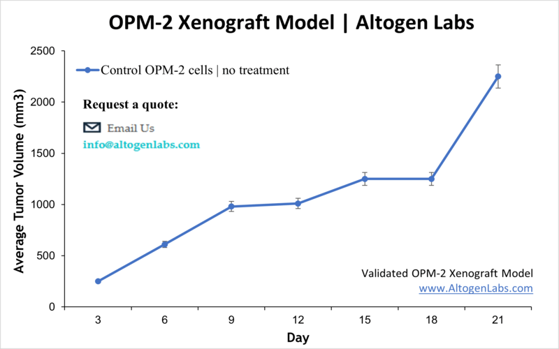
- End of study is reached when tumor size reaches 2,000 mm3 or the predetermined size limit per approved IACUC protocol.
- Final necropsy and tissue collections are performed for appropriate downstream analysis. Tumors are excised, weighed and documented by digital imaging. Tumors and tissues can be stabilized in RNAlater reagent, snap frozen in LN2 or prepared for histology.