Hepa 1-6 Allograft Syngeneic model
Hepa1-6 is a murine hepatoma cell line that is widely used in cancer research as a model system to study liver cancer biology. It was originally derived from a spontaneous hepatoma in a C57BL/6 mouse and has been extensively characterized for its growth properties, gene expression profile, and response to various anti-cancer agents. One of the advantages of the Hepa1-6 cell line is its ability to form tumors when transplanted into immunocompetent syngeneic mice, which allows researchers to study the interaction between the tumor and the immune system in a more physiologically relevant setting. This feature has made Hepa1-6 a valuable tool for studying the role of the immune system in tumor growth and response to therapy.
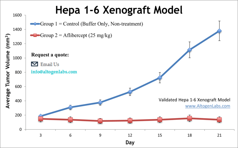
Hepatocellular carcinoma (HCC) is a significant cause of cancer mortality worldwide. Liver cancer is often associated with a poor prognosis, and the exact mechanisms leading to its development are still unclear. The Hepa 1-6 tumorigenic epithelial cell line is derived from liver cells of the BW7756 mouse (Mus musculus) with hepatoma. Hepa 1-6 has been negatively tested for mousepox and is widely used in mammalian liver tissue functions research. A 2013 study in PLoS demonstrates that Hydroxysteroid sulfotransferase 2B1b SULT2B1 is overexpressed in the human hepatocarcinoma tumorous tissues compared to their adjacent tissues, which promotes the growth of the mouse hepatocarcinoma cell line Hepa1-6. Also, knock-down of SULT2B1b inhibits cell growth and cyclinB1 levels in human hepatocarcinoma cells and suppresses xenograft growth in vivo. A 2017 study (Nevzorova et a.) used the Hepa-1-6 model to test for both in vitro and in vivo effects of Abisilin®, a natural novel pharmacological terpenoid compound originally extracted from coniferous Pinaceae tress; results demonstrated the compound has anti-angiogenic and anti-tumor properties in terms of growth, apoptosis, proliferation and vasculature. Kim et al. also released a study (2015) using Hepa-1-6 cells to examine cancer-specific post-transcriptional targeting as a method of cancer therapy. Tey developed trans-splicing ribozomes of Tetrahymena group I intron in order to target RNA of HCC specific telomerase reverse transcriptase (TERT); adenovirus transfection with these ribozymes showed low-toxicity anti-tumor effects which holds potential for a safe and effective clinical targeting strategy. Finally, a 2017 study released by Chuang et al. used the Hepa 1-6 xenograft model to characterize JC-001, a Chinese medicine traditionally used for non-cancer liver disease such as chronic hepatitis C and B, in tumors. Results suggested an immunomodulation effect with JC-001 treatment as there was increased immune cell infiltration as well as increased expression of Ki67 and hypoxia inducible factor 1 alpha (HIF-1α), providing support for the use of JC-001 in clinical applications of hepatoma treatment. The Hepa 1-6 cell line (mouse liver) is used to create the Cell Line Derived (CDX) xenograft Hepa 1-6 mouse model. The Hepa 1-6 xenograft model enables the study of tumor growth inhibition of therapeutic agents such as expression of hTERTC27 by a recombinant adenovirus or RNA replacement therapy through miRNA regulation (i.e. miR-122 or miR-34).
Download Altogen Labs Hepa1-6 Xenograft Model PowerPoint Presentation: ![]()
Basic study design
- Exponential growth of cells is maintained prior to collection.
- The cells are collected for inoculation by trypsinizing the cells in the flasks. Cell count and viability is then determined following a flow cytometry or trypan blue exclusion test (98% minimum viability). Suspensions are then adjusted to the density required for inoculation.
- One million cells, in a volume of 100-200 µL of the Matrigel + Hepa 1-6 cells suspension, is inoculated subcutaneously (s.c) into the hind leg of each mouse (NOD/SCID or athymic BALB/C, 11-12 weeks).
- The injection sites are observed until palpated tumors are established. Calipers are used for measuring tumors until average sizes of 90-140 cubic mm are reached.
- After sorting (randomization) into appropriate treatment cohorts, the test compounds are administered according to an agreed upon treatment schedule.
- Daily tumor measurements are recorded and mouse weights are documented 3 times weekly.
- As the study’s tumor size limit is reached (or 2,000 cubic mm), the animals are euthanized humanely.
- As predetermined for the end of the study, a necropsy is performed.
- The tumors are removed, their weight recorded, and then the tumor is documented using digital imaging.
- Predetermined tissues are collected (snap frozen, submersed in RNALater reagent, isolation of nucleic acids or prepared for histological analysis) by performing standard gross necropsies.
Get Instant Quote for
Hepa1-6 Xenograft Model
Altogen Labs provides an array of laboratory services using over 90 standard Cell Line Derived Xenograft models and over 30 PDX models. We provide quantitative gene expression analysis of mRNA expression (qPCR) and protein expression analysis using the WES system (ProteinSimple). Animal handling and maintenance at the Altogen Labs facilities are IACUC regulated and GLP-compliant. Following acclimatization to the vivarium environment, mice are sorted according to body mass. The animals are examined daily for tumor appearance and clinical signs. We provide detailed experimental procedures, health reports and data (all-inclusive report is provided to the client that includes methods, results, discussion and raw data along with statistical analysis). Additional services available include collection of tissue, histology, isolation of total protein or RNA and analysis of gene expression.
Following options are available for the Hepa-1-6 xenograft model:
- Hepa 1-6 Tumor Growth Delay (TGD; latency)
- Hepa 1-6 Tumor Growth Inhibition (TGI)
- Dosing frequency and duration of dose administration
- Dosing route (intravenous, intratracheal, continuous infusion, intraperitoneal, intratumoral, oral gavage, topical, intramuscular, subcutaneous, intranasal, using cutting-edge micro-injection techniques and pump-controlled IV injection)
- Immunohistochemistry, lipid distribution and metabolic assays
- Imaging studies: Fluorescence-based whole body imaging
- Alternative cell engraftment sites (orthotopic transplantation, tail vein injection and left ventricular injection for metastasis studies, injection into the mammary fat pad, intraperitoneal injection)
- Blood chemistry analysis
- Toxicity and survival (optional: performing a broad health observation program)
- Gross necropsies and histopathology
- Positive control group employing a known chemotherapy, usually at a dosage of 10-20 mg/kg
