
WiDr xenograft model
The WiDr cell line is a human colorectal adenocarcinoma model that was originally derived in 1971 from the tumor of a 44-year-old female patient. It is widely used in oncology research for its consistent tumorigenic capacity in immunodeficient mice and well-characterized molecular features. Although historically related to the HT-29 line, WiDr possesses distinct biological characteristics, including expression of carcinoembryonic antigen (CEA), colon-specific antigen (CSAp), epidermal growth factor (EGF), and transforming growth factor beta (TGF-β). The cell line is also keratin-positive by immunoperoxidase staining, expresses p53 antigen, and is negative for colon antigen 3. These traits contribute to its reliability in modeling epithelial tumor behavior, therapeutic responses, and tumor-stroma interactions in xenograft systems. WiDr xenograft models have been employed extensively in the evaluation of antineoplastic agents targeting a range of molecular pathways. A landmark study by Shindoh et al. demonstrated that the novel histone deacetylase (HDAC) inhibitor YM753 induced tumor-selective cytotoxicity, accompanied by tumor growth suppression and histone hyperacetylation within the tumor tissue, but not in normal cells. Pharmacokinetic analyses revealed that the reduced form of YM753 accumulated selectively in tumor tissues, despite being rapidly cleared from plasma, indicating potential for tumor-specific retention and clinical utility. Similarly, a comparative study investigating the semisynthetic camptothecin derivative CPT-11 and its active metabolite SN-38 established that the activity of DNA topoisomerase I, rather than carboxylesterase expression, was a key determinant of drug sensitivity. These findings not only underscored the clinical efficacy of CPT-11, now marketed as irinotecan, but also validated WiDr xenografts as a predictive model for response to topoisomerase-targeting therapies. Further highlighting its versatility, the WiDr model has been employed in anti-angiogenic therapy research. A study using the receptor tyrosine kinase inhibitor TSU-68 (SU-6668) demonstrated significant in vivo inhibition of tumor growth and metastasis by disrupting VEGF, basic fibroblast growth factor (bFGF), and platelet-derived growth factor (PDGF) signaling. The high endogenous expression of epidermal growth factor receptor (EGFR) in WiDr cells makes this model particularly advantageous for investigating EGFR-targeted therapies and their combinatorial use with other agents such as irinotecan or gefitinib. WiDr xenografts serve as an ideal platform for assessing the pharmacodynamics of EGFR tyrosine kinase inhibitors (EGFR-TKIs) and for validating therapeutic strategies that combine molecular pathway inhibitors with conventional chemotherapeutics. Altogen Labs utilizes the WiDr cell line to establish reliable cell line-derived xenograft (CDX) models, supporting preclinical studies on epigenetic modulation, DNA replication inhibition, and receptor-mediated signaling blockade. With its reproducible tumor growth kinetics, defined antigenic and molecular signature, and responsiveness to a wide spectrum of pharmacologic agents, the WiDr xenograft model remains a valuable tool for translational colorectal cancer research.
WiDr Subcutaneous and Orthotopic Xenograft Models: Download ![]()
Download Altogen Labs WiDr Xenograft Model PowerPoint Presentation: ![]()
WiDr Cell Line
The WiDr cell line, derived from a human colorectal adenocarcinoma, is widely employed as a model system in colorectal cancer research due to its well-characterized genetic and molecular profile. It shares a common origin with the HT-29 cell line and displays epithelial morphology, along with a mutant TP53 gene and deregulated signaling through the Wnt/β-catenin and EGFR pathways. WiDr cells are KRAS wild-type but exhibit elevated expression of EGFR and HER2, making them suitable for evaluating targeted therapies such as cetuximab, although responses are often limited by mechanisms of therapeutic resistance that remain incompletely understood. Additionally, this cell line expresses high levels of COX-2 and has been utilized in studies investigating the chemopreventive potential of COX-2 inhibitors and nonsteroidal anti-inflammatory drugs. Recent high-throughput analyses have further implicated dysregulation in MAPK/ERK signaling, mitochondrial bioenergetics, and lipid metabolism as contributors to tumor progression and drug resistance.
WiDr Subcutaneous Xenografts in Colorectal Cancer Modeling
Subcutaneous xenograft transplantation is a widely used method for modeling human tumor growth in vivo and remains a foundational approach in preclinical colorectal cancer research. In this model, WiDr colorectal adenocarcinoma cells are implanted into the subcutaneous space of immunocompromised mice, where they form measurable tumors that support controlled evaluation of therapeutic efficacy. The WiDr cell line, characterized by epithelial morphology, mutant TP53, wild-type KRAS, and deregulated Wnt/β-catenin and EGFR signaling, provides a reproducible and biologically relevant model for assessing the activity of targeted therapies and chemotherapeutics. The subcutaneous site offers ease of tumor monitoring through external caliper measurements, permitting frequent, non-invasive assessments of tumor progression and drug response throughout the study duration.
Beyond its procedural accessibility, the WiDr xenograft model supports detailed downstream analyses, including tumor weight quantification, histological examination, and molecular profiling of treatment-induced changes in cell signaling, apoptosis, and proliferation. The model is compatible with various dosing routes and regimens, allowing for comprehensive evaluation of single agents and combinatorial strategies. Although limited in its ability to replicate the tumor microenvironment and immune contexture, the WiDr subcutaneous xenograft remains a critical platform for screening drug candidates, optimizing therapeutic schedules, and generating mechanistic insights. Its integration into colorectal cancer research continues to provide a robust and scalable framework for advancing the development of more effective and personalized treatment strategies.
Modeling Colorectal Metastasis with WiDr Xenografts
Metastatic xenograft transplantation serves as a critical platform for modeling tumor dissemination and evaluating therapeutic strategies aimed at preventing or controlling metastatic progression in colorectal cancer. While the WiDr cell line is traditionally employed in subcutaneous xenograft studies, it has also demonstrated the capacity to form metastases in vivo, offering unique opportunities to study advanced disease states. Specifically, WiDr-derived metastases have been established in immunodeficient mice, most notably in the liver following intrasplenic or portal vein injection, thereby replicating a clinically relevant pattern of colorectal cancer spread. These models allow for controlled examination of metastatic colonization, growth kinetics, and the impact of systemic therapies on established secondary lesions.
The ability of WiDr cells to metastasize in vivo reflects their intrinsic molecular features, including aberrant EGFR signaling, impaired TP53 function, and upregulation of pro-invasive mediators such as COX-2. Metastatic xenograft models using WiDr cells enable rigorous investigation into the molecular and cellular mechanisms governing metastatic seeding, organotropism, and therapeutic resistance. Such models are particularly valuable for evaluating agents that may target microenvironmental interactions, matrix remodeling, angiogenesis, and immune evasion mechanisms that are difficult to study in conventional subcutaneous settings. Although challenges remain in replicating the full complexity of metastatic progression, WiDr-based metastatic xenografts enhance the translational value of preclinical research by providing a system in which to test anti-metastatic agents, dissect mechanisms of treatment escape, and identify biomarkers predictive of distant spread. These models thus represent an essential extension of xenograft methodology, supporting the broader objective of refining cancer therapeutics through biologically relevant in vivo systems.
Metabolic Modulation of Chemotherapy Response in WiDr Cells
WiDr is a human colorectal adenocarcinoma cell line commonly used to examine chemotherapy response and the influence of metabolic conditions on tumor cell behavior. It carries mutations in TP53, PIK3CA, and BRAF, which contribute to deregulated proliferation and altered intracellular signaling through pathways such as PI3K/AKT and MAPK. When cultured in elevated levels of insulin, IGF-1, and glucose, WiDr cells exhibit increased metabolic activity and cell viability. This effect is particularly notable under high glucose conditions combined with IGF-1 exposure, suggesting that survival pathways are enhanced through insulin-like signaling under metabolically rich environments. Insulin demonstrates more variable effects, occasionally enhancing drug sensitivity depending on glucose concentration and exposure duration. WiDr cells show distinct alterations in drug response when exposed to chemotherapy agents such as 5-fluorouracil and oxaliplatin following metabolic pretreatment. IGF-1 tends to increase resistance, potentially by activating anti-apoptotic pathways, while insulin can either sensitize the cells or produce no significant effect. Interestingly, the addition of ultra-low concentrations of chemotherapy in combination with IGF-1 can increase WiDr cell viability above baseline, indicating a potentially adverse interaction between subtherapeutic drug levels and growth-promoting signals.
IGF-1 and Insulin Modulate Drug Sensitivity in WiDr Cells
WiDr is a human colorectal adenocarcinoma cell line often used to investigate cellular responses to chemotherapy and the influence of metabolic and hormonal conditions on tumor behavior. When WiDr cells are exposed to increased concentrations of insulin and insulin-like growth factor-1 (IGF-1), they exhibit enhanced viability and metabolic activity, particularly under high-glucose conditions. IGF-1 appears to stimulate resistance to commonly used chemotherapeutic agents such as oxaliplatin and 5-fluorouracil, especially when cells are pretreated with the growth factor in a normal-glucose environment. In contrast, insulin exposure under both glucose conditions generally sensitizes WiDr cells to chemotherapy, reducing cell viability. The apparent upregulation of survival-promoting pathways, potentially mediated by IGF-1 receptor activation and modest increases in HIF-1α expression, contributes to altered sensitivity.
WiDr as a Model for MAPK-Driven Colorectal Cancer
The WiDr colon carcinoma cell line demonstrates a complex oncogenic profile that reflects its utility as a model for studying colorectal cancer progression and therapeutic response. Genomic analysis of WiDr reveals key mutations in several driver oncogenes, most notably BRAF V600E, which promotes constitutive MAPK pathway activation and drives proliferation independent of upstream EGFR signaling. This mutation renders the cells relatively unresponsive to EGFR-targeted therapies, a resistance mechanism reinforced by concurrent activation of downstream effectors such as MEK and ERK. In addition, WiDr lacks mutations in KRAS and PIK3CA, distinguishing it from many other colorectal cancer cell lines and allowing clearer attribution of signaling behavior to the BRAF mutation. While APC and TP53 are commonly mutated in colorectal cancer, WiDr displays wild-type TP53 status, which may affect responses to DNA-damaging agents and alter apoptotic sensitivity. Experimental findings show that WiDr cells exhibit sustained ERK phosphorylation and high levels of cyclin D1, consistent with hyperactive mitogenic signaling. Therapeutic inhibition of BRAF or MEK induces partial growth suppression, but residual signaling often persists, indicating pathway reactivation or compensatory feedback. These observations support the hypothesis that BRAF V600E acts as a dominant oncogenic driver in WiDr, while other signaling networks modulate therapeutic efficacy.
WiDr Subcutaneous and Orthotopic Xenograft Models: Download ![]()
Basic study design
- WiDr cells are collected with trypsin-EDTA. Viable cells are counted using trypan blue exclusion. The mice (athymic BALB/C or NOD/SCID, 11 to 13 weeks) are injected subcutaneously in the flank of a hind leg. One million cells are inoculated into the mice (vol = 140-180 microliters of matrigel plus WiDr cells suspension).
- The injection are continuously observed until tumors are established. Tumors are measured until they reach an average of 100-150 mm3. Animals are sorted into treatment cohorts and the in-life portion of the study begins.
- Injections of test material is performed following the dosing schedule. Tumors are continually measured and body weights recorded.
- At the end of the study necropsies and tissue collections are followed according to the study design. Tumors are excised and weighed. All tissues collected can be placed in RNA-Later, snap frozen or added to 10% NBF for histology.
Get Instant Quote for
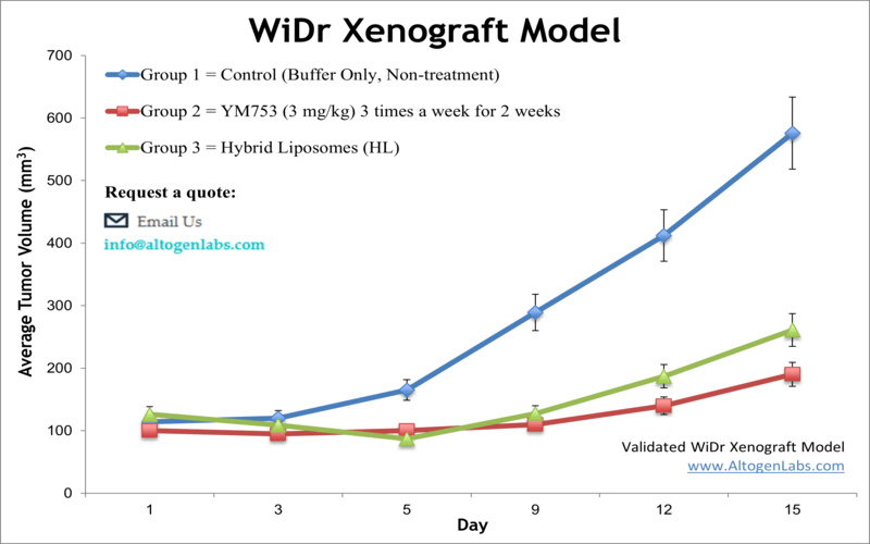
WiDr Xenograft Model
Xenograft animal models are used to assess the effectiveness of drugs against specific types of cancer. New medicines are tested on staged tumor growths that have been engrafted via subcutaneous or orthotopic inoculation in an immunocompromised mouse or rat model. All clinically approved anti-cancer agents have been evaluated with conventional preclinical in vivo models. Xenograft studies can be highly complex, starting with the selection of the appropriate animal model, choice of tumorigenic cell line, administration method, dosing, analysis of tumor growth rates and tumor analysis (histology, mRNA and protein expression levels). Animal handling and maintenance at the Altogen Labs facility is IACUC regulated and GLP compliant. Following acclimation to the vivarium environment, mice are sorted according to body mass. The animals are examined daily for tumor appearance and clinical signs. We provide detailed experimental procedures, health reports and data (all-inclusive report is provided to the client that includes methods, results, discussion and raw data along with statistical analysis).
Following options are available for the WiDr xenograft model:
- WiDr Tumor Growth Delay (TGD; latency)
- WiDr Tumor Growth Inhibition (TGI)
- Dosing frequency and duration of dose administration
- Dosing route
- WiDr tumor immunohistochemistry